
follows rigorous standards of quality and accountability. is accredited by URAC, for Health Content Provider (URAC's accreditation program is an independent audit to verify that A.D.A.M. Lying on the back with the thighs and legs flexed and turned (lithotomy position) during surgery or diagnostic proceduresĪ.D.A.M., Inc.Internal bleeding in the pelvis or belly area (abdomen).Diabetes or other causes of peripheral neuropathy.A catheter placed into the femoral artery in the groin.The femoral nerve can also be damaged from any of the following: Compression, stretching, or entrapment of the nerve by nearby parts of the body or disease-related structures (such as a tumor or abnormal blood vessel).More common causes of femoral nerve dysfunction are: Disorders that involve the entire body ( systemic disorders) can also cause isolated nerve damage to one nerve at a time (such as occurs with mononeuritis multiplex). Mononeuropathy usually means there is a local cause of damage to a single nerve. It provides feeling (sensation) to the front of the thigh and part of the lower leg.Ī nerve is made up of many fibers, called axons, surrounded by insulation, called the myelin sheath.ĭamage to any one nerve, such as the femoral nerve, is called mononeuropathy. It helps the muscles move the hip and straighten the leg. All rights reserved.The femoral nerve is located in the pelvis and goes down the front of the leg. Athletes with previous groin injury had a significant fall in some EMG outputs.Ĭopyright © 2011 Elsevier Ltd. Muscle EMG varied significantly with clinical test position. Hip adduction strength assessment is best measured at hips 0 (which produced most force) or 45° flexion (which generally gave the highest EMG output).

All other factors had no significant effect on the force outputs.

BMI (body mass index) was a significant factor (p < 0.01) for producing a higher force. For force data, clinical test type was a significant factor (p < 0.01) with Hips 0 being significantly stronger than Hips 45, Hips 90 and Side lay. Injury history was a significant factor in the EMG output for the adductor longus (p < 0.05), pectineus (p < 0.01) and gracilis (p < 0.01) but not adductor magnus. EMG activation for pectineus was highest in Hips 90. EMG activation was highest in Hips 0 or Hips 45 for adductor magnus, adductor longus and gracilis. Test type was a significant factor in the EMG output for all four muscles (all muscles p < 0.01). A load cell was used to measure force data.

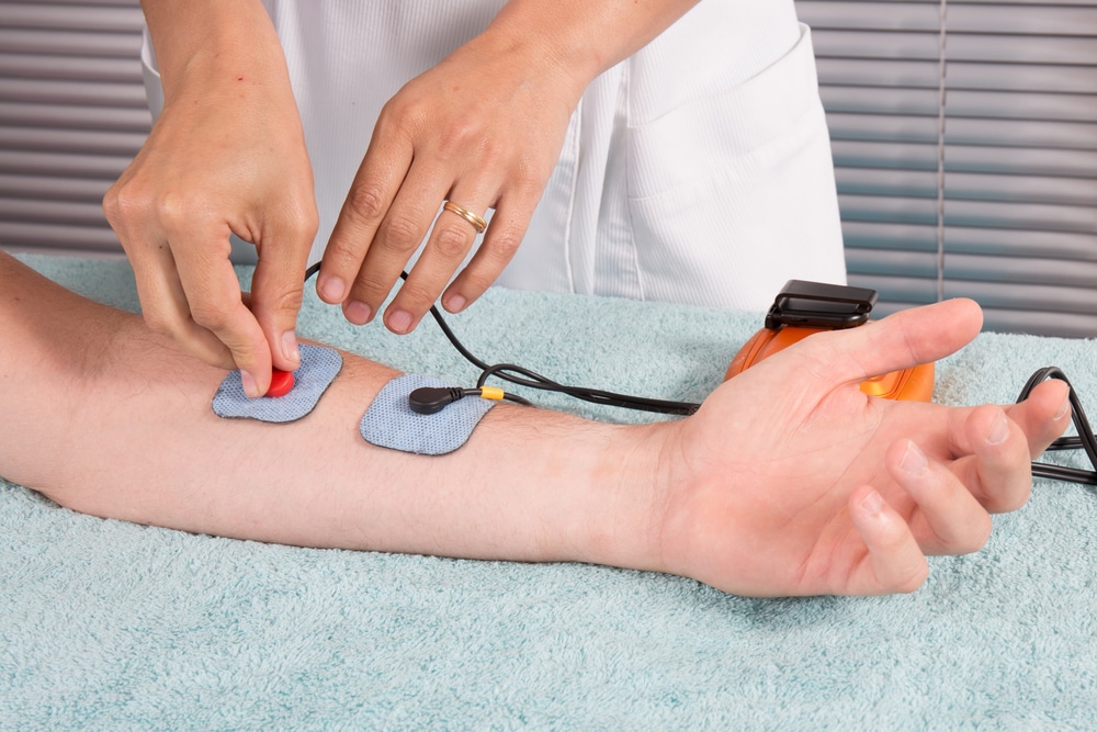
To assess activation of muscles of hip adduction using EMG and force analysis during standard clinical tests, and compare athletes with and without a prior history of groin pain.Ģ1 male athletes from an elite junior soccer program.īilateral surface EMG recordings of the adductor magnus, adductor longus, gracilis and pectineus as well as a unilateral fine-wire EMG of the pectineus were made during isometric holds in six clinical examination tests.


 0 kommentar(er)
